Compiled from public reports
Why a team treated myasthenia gravis before removing a thymoma
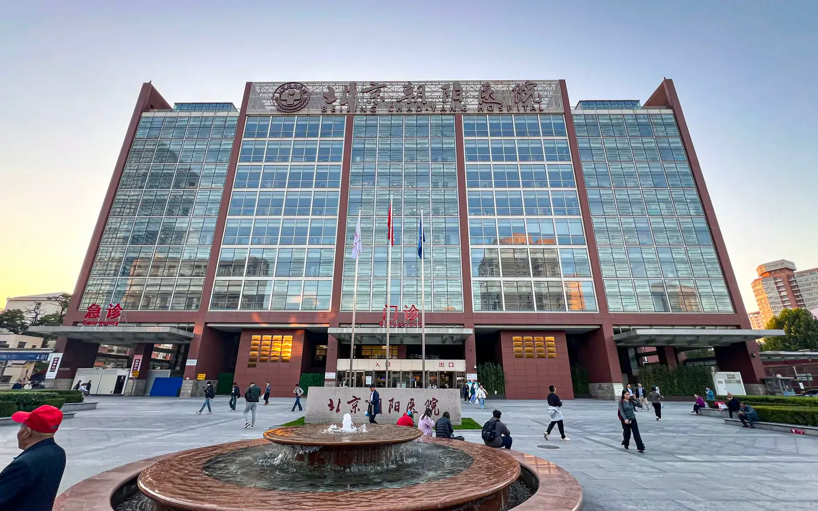
When chest imaging revealed a mass in the thymus, the patient already had difficulty swallowing and shortness of breath. Beijing Chaoyang Hospital treated the myasthenia gravis before removing the mass once the symptoms were controlled.
Compiled by SinoCareHub · Information checked: 2026-11-07
Patient overview
An adult patient · China · Beijing · Medical treatment followed by thymoma removal
- Treatment in China:
- The team treated the myasthenia gravis first, then removed the thymoma and monitored the patient in intensive care.
- Outcome at the reported stage:
- Swallowing improved before discharge. The hospital recorded no myasthenic crisis during that stay and did not report longer-term follow-up.
From limb weakness to choking when drinking
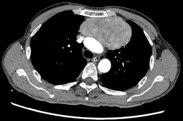
At first the patient felt weak in his arms and legs and tired more easily with activity. His symptoms were milder in the morning, worse in the evening and improved with rest. As they progressed, he began choking when drinking water, had trouble swallowing and became short of breath after activity. A chest CT scan showed a mass behind the breastbone in the front part of the space between the lungs. He went to the neurology department at Beijing Chaoyang Hospital for further evaluation.
Why the team did not remove the mass immediately
Qin Wei’s team diagnosed generalized myasthenia gravis and a thymoma more than 10 cm across. The mass was close to major blood vessels and the nerves that help control the diaphragm. The patient’s breathing and nutritional status added to the surgical risk. The team paid particular attention to the risk of a myasthenic crisis around the time of surgery, which could interfere with breathing.
The neurology team started immunotherapy. A CT-guided needle biopsy through the skin and a PET-CT scan helped assess the thymic lesion. The team also assessed lung function and addressed nutritional risks. Thoracic surgery, anaesthesia and intensive care staff drew up a plan for the period around surgery. Surgery was scheduled once the symptoms of myasthenia were under control.
Intensive care monitoring followed open chest surgery
The teams of Hu Bin and Chen Shuo operated through the centre of the breastbone. They separated the mass from nearby blood vessels and the phrenic nerves, then removed it along with fatty tissue in the front part of the space between the lungs. The hospital reported that surgery lasted about four hours. The patient then went to intensive care for monitoring.
Swallowing improved before discharge
According to the hospital, the patient did not develop a myasthenic crisis during this hospital stay. Treatment continued after he returned to neurology. Before discharge, strength in muscles closer to the trunk was grade 4 and strength farther out in the limbs was grade 5. Swallowing had also improved markedly. The report ended at discharge and gave no longer-term update on myasthenia symptoms or follow-up for the tumour.
Plan care in China
Ask about medical records and contacting hospitals.