
Visit preparation
Why test for glaucoma when eye pressure is normal?
A single eye pressure reading in the normal range can still warrant testing for glaucoma. If a report from an examination of the back of the eye flags a possible problem with the optic nerve, the doctor compares how the nerve looks with how well you see across your visual field before deciding whether to monitor, run more tests, or make a diagnosis.
Eye pressure and the optic nerve tell you different things
An eye pressure test measures the pressure inside your eye at that moment. To assess for glaucoma, the doctor also checks whether the optic nerve is damaged and whether that damage affects the area you can see. Normal-tension glaucoma is a type of open-angle glaucoma in which characteristic optic nerve damage occurs even when eye pressure is within the usual reference range. Early on, you may still read or see your phone screen clearly. An eye chart mainly measures central visual acuity and cannot replace a visual field test. [1,2]
A checkup report that notes a “large cup-to-disc ratio” is describing the shape of the optic disc, the visible head of the optic nerve. That finding alone cannot establish a diagnosis. Some people naturally have larger optic discs, and high myopia (severe nearsightedness) can make the results harder to interpret. A “normal” eye pressure reading likewise does not rule out a suspicious finding in the optic nerve. [1,2]
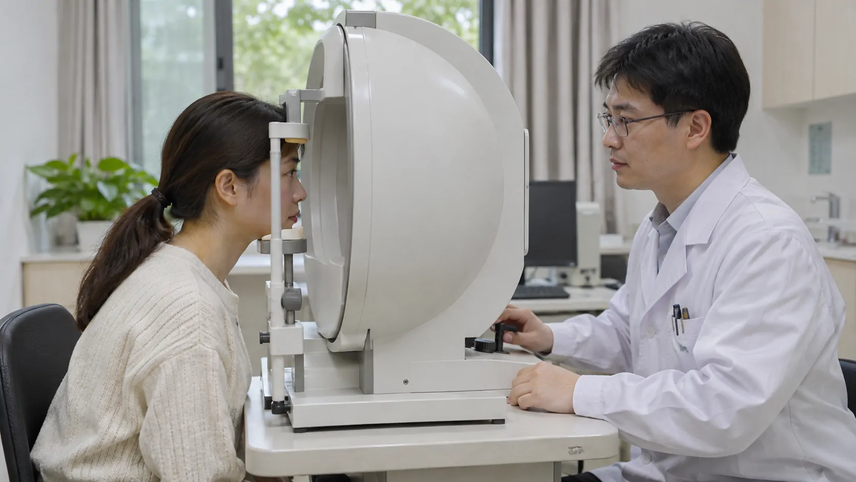
Why do both an OCT scan and a visual field test?
- An optic nerve OCT scan shows structure. Optical coherence tomography (OCT) helps show whether the retinal nerve fibre layer around the optic disc has thinned. The colours on the scan show how the results compare with the device’s reference database. The doctor still needs to review the original images and check their quality.
- A visual field test measures function. It records where you tend to miss spots of light. If attention or difficulty following the test instructions affects the first result, the doctor may repeat the test to see whether the same defect appears again.
- Drainage angle and eye pressure tests help identify the type of glaucoma. The doctor examines the angle through which fluid drains from the eye. As needed, they consider eye pressure readings taken at different times and other information such as corneal thickness to interpret what a single measurement might miss. [1]
What matters is whether the results point to the same problem. For example, the doctor compares thinning at a particular part of the optic nerve with the location of a visual field defect in the same eye. If only one result is abnormal, the doctor may first establish a baseline that can be compared with later tests to see whether the finding changes over time. [1]
Which reports should you bring to a follow-up in China?
When visiting an ophthalmology clinic with a glaucoma specialist, arrange your checkup report, earlier photos of the back of the eye, optic nerve OCT scans, and visual field charts by date. Label each with the right or left eye. If you have used eye drops, write down their names and how you used them. At the visit, you can ask, “Does the optic nerve only look suspicious, or do the structural and visual field findings already match? Which test should we use for comparison next time?” This helps clarify whether the next step is to confirm the diagnosis or to monitor progression. [1,2]
If the doctor confirms normal-tension glaucoma, they may still discuss lowering eye pressure further to protect the optic nerve. The specialist will set your target eye pressure, treatment, and follow-up schedule based on how much damage there is and how it changes. If you already plan to see a doctor at another hospital, see the guide to preparing medical records.
Need help?
Tell us about your situation and needs. Incomplete records are welcome.
Related reading
Sources and references
- Glaucoma Group, Chinese Ophthalmological Society, Chinese Medical Association. Chinese expert consensus on the diagnosis and treatment of normal-tension glaucoma (2019), May 2019. · 2019-05
- Beijing Friendship Hospital. A guide to glaucoma prevention, diagnosis, and treatment, 7 March 2025. · 2025-03-07