
Visit preparation
Why might a small kidney stone still need treatment?
Size alone does not determine treatment. A small stone remaining in the kidney differs from one that enters the ureter, obstructs urine or accompanies infection. “Minimally invasive” also describes different procedures.
A few millimetres do not determine risk
A small kidney stone in an adult may cause no symptoms, or may block urine flow after moving. Decisions depend not only on size but also location, drainage, infection and possible impairment of kidney function.[1,2]
Urine passes from the kidneys through the ureters to the bladder. A stone in the kidney and one in the ureter cannot be compared by diameter alone. Beijing Friendship Hospital’s urology information explains that stones only a few millimetres across can lodge in the urinary tract and cause obstruction, pain or kidney damage.[1]
Small does not mean definitely safe. Conversely, finding a stone on a check-up does not automatically require immediate removal. Doctors may consider observation for small kidney stones without obstruction or infection, depending on the person.[1]
What observation needs to assess
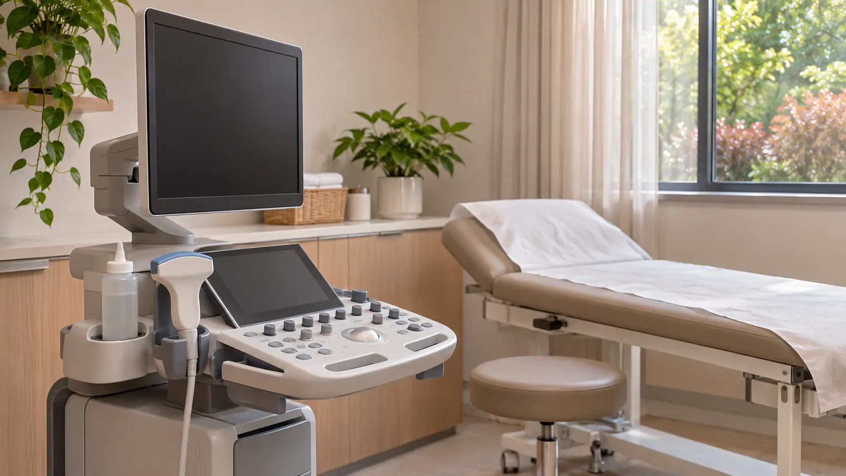
Observation still involves understanding location and later changes. Depending on the situation, ultrasound, CT, urine and blood tests can help assess obstruction, infection and other problems.[1,2]
Beijing Friendship Hospital explains that ultrasound can show stones and urine-related dilation, while CT helps locate stones and assess obstruction.[1] Words such as “ureter” or “hydronephrosis”—dilation of the kidney’s collecting system—may therefore clarify the problem better than size alone. The doctor decides whether existing information needs further tests.
Seek prompt medical care for severe pain, fever or chills when a stone is suspected.[2] Pain with fever should not simply be explained as a stone moving out. Do not wait for an appointment in China or a platform response.
Different routes for breaking up stones
If treatment is needed, common options include extracorporeal shock-wave lithotripsy, ureteroscopy and percutaneous nephrolithotomy. These are different procedures.[1,2]
Shock waves applied outside the body break a stone into fragments that pass through the urinary tract. Ureteroscopy passes through the urethra and bladder into the ureter or kidney. Percutaneous nephrolithotomy creates a passage through the back into the kidney.[1]
When comparing plans in China, identify the exact procedure and why it fits the stone’s location and circumstances before discussing burdens and risks. “Laser” does not necessarily mean no incision; “minimally invasive” does not mean an identical route. Some endoscopic procedures require general anaesthesia. The urology and relevant teams should explain anaesthesia, individual risks and follow-up.[2]
Stone composition still matters
Stones have different compositions. Beijing Friendship Hospital distinguishes calcium-containing, uric acid and infection-related stones, among others. Analysis of a passed or removed stone may help guide more targeted prevention.[1]
This is why one food-avoidance list cannot simply fit everyone. Keep stone analysis, imaging and operation records so the next doctor knows what was treated and what still needs observation. See the return-home follow-up guide for care across hospitals or countries.
This article does not prescribe self-directed stone passage, medicines or changes to fluid intake. A doctor should determine whether to observe or intervene, which procedure to use and how to reduce recurrence.
Need help?
Tell us about your situation and needs. Incomplete records are welcome.