
Visit preparation
Why might I need OCT after a diabetic eye screening photo?
Retinal photos can show bleeding and deposits called exudates on the retinal surface. If the eye doctor suspects macular edema, they may add an OCT scan to see whether thickening or fluid affects the part of the macula responsible for central vision.
What does each test show?
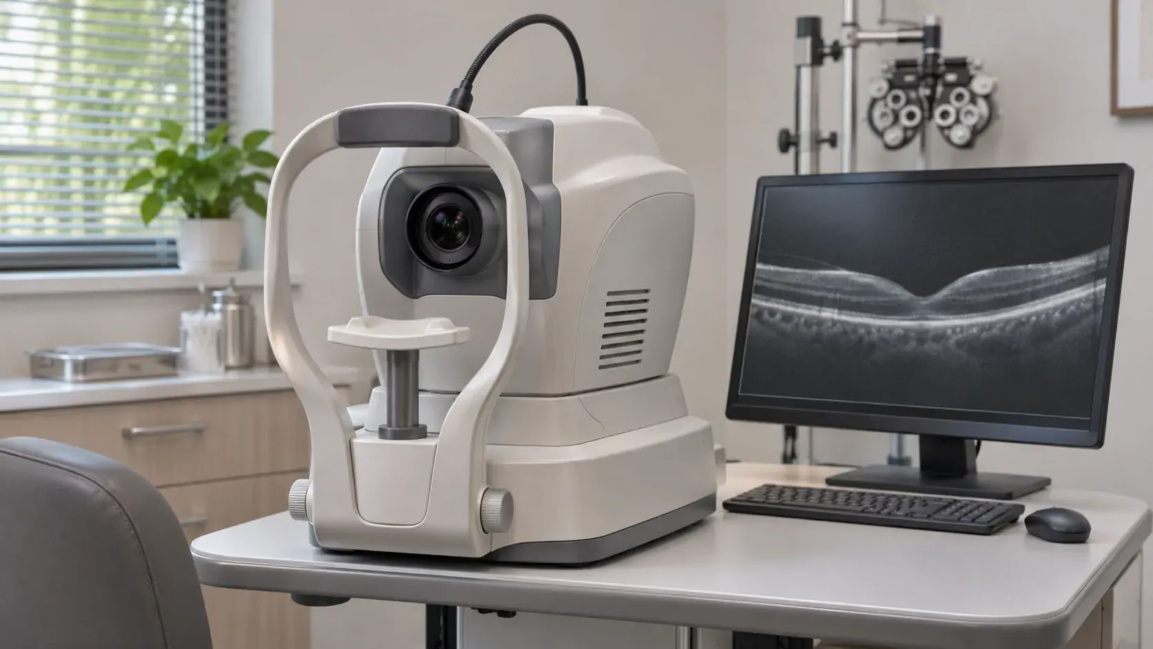
Retinal photography mainly shows the surface of the retina and can reveal changes such as bleeding and deposits called exudates. Optical coherence tomography (OCT) provides cross-sectional images of the macula. These help the eye doctor see whether the retina has thickened or fluid has collected, and whether the center of the macula is affected. [1,2]
The center of the macula helps you see fine detail when reading or recognizing faces. If your report gives a diabetic retinopathy grade, it is describing how severe the changes at the back of the eye are. Whether there is macular edema and whether it affects the center are separate findings that can influence the next assessment. One report can answer both questions, and your doctor may need both tests to do so. [1,2]
Check which eye and which finding the report describes
- “No abnormality seen” in one eye. Check which eye and which test this refers to. Read the result for the other eye separately. [2,3]
- “Poor image quality” or “ungradable.” The photos are insufficient to make an assessment. Bring the original images to the eye doctor, who can decide whether to repeat the photos or examine your eyes after dilating the pupils. [2,3]
- “Suspected macular edema.” The eye doctor can review your visual acuity, examine the back of the eye, and use OCT to check whether cross-sectional images show thickening or fluid. Whether you need OCT depends on what the doctor needs to confirm. [1,2]
What does the eye doctor check after screening?
China's diabetes prevention and care guideline lists retinal photography without pupil dilation as a tool endocrinology clinics can use for screening and follow-up. If there are signs of a problem, the eye doctor can take photos with the pupils dilated or examine the back of the eye with a slit lamp, among other approaches, to make a diagnosis and grade the changes. OCT primarily helps assess macular edema. The eye doctor still needs to examine areas beyond the macula and consider other findings. [1,2]
If you are referred to an eye doctor, you can ask, “Am I being referred because the photos were unclear, because they showed changes, or because the doctor needs to check for macular edema?” The reason helps determine what the eye doctor needs to check. [1–3]
Compare results for the same eye at follow-up
Keep the original retinal photos, OCT images, and reports for both eyes, and date each item. If you go to two clinics or hospitals, ask the doctor to compare visual acuity and cross-sectional images of the macula for the same eye first. Then ask which changes need further monitoring. At your first eye appointment, the doctor will also ask how long you have had diabetes, whether you have had any eye treatment, and how well your blood sugar has been controlled recently. Bring any records you have. [1,2]
If your vision suddenly worsens noticeably, seek urgent assessment at a nearby eye emergency service. [1,2]
Need help?
Tell us about your situation and needs. Incomplete records are welcome.